Osmium »
PDB 1hc8-8uiw »
6hzl »
Osmium in PDB 6hzl: Crystal Structure of Redox-Inhibited Phosphoribulokinase From Synechococcus Sp. (Strain Pcc 6301), Osmate Derivative
Enzymatic activity of Crystal Structure of Redox-Inhibited Phosphoribulokinase From Synechococcus Sp. (Strain Pcc 6301), Osmate Derivative
All present enzymatic activity of Crystal Structure of Redox-Inhibited Phosphoribulokinase From Synechococcus Sp. (Strain Pcc 6301), Osmate Derivative:
2.7.1.19;
2.7.1.19;
Protein crystallography data
The structure of Crystal Structure of Redox-Inhibited Phosphoribulokinase From Synechococcus Sp. (Strain Pcc 6301), Osmate Derivative, PDB code: 6hzl
was solved by
R.H.Wilson,
A.Bracher,
F.U.Hartl,
M.Hayer-Hartl,
with X-Ray Crystallography technique. A brief refinement statistics is given in the table below:
| Resolution Low / High (Å) | 30.00 / 2.77 |
| Space group | H 3 2 |
| Cell size a, b, c (Å), α, β, γ (°) | 141.432, 141.432, 206.563, 90.00, 90.00, 120.00 |
| R / Rfree (%) | 23.5 / 25.9 |
Osmium Binding Sites:
The binding sites of Osmium atom in the Crystal Structure of Redox-Inhibited Phosphoribulokinase From Synechococcus Sp. (Strain Pcc 6301), Osmate Derivative
(pdb code 6hzl). This binding sites where shown within
5.0 Angstroms radius around Osmium atom.
In total 2 binding sites of Osmium where determined in the Crystal Structure of Redox-Inhibited Phosphoribulokinase From Synechococcus Sp. (Strain Pcc 6301), Osmate Derivative, PDB code: 6hzl:
Jump to Osmium binding site number: 1; 2;
In total 2 binding sites of Osmium where determined in the Crystal Structure of Redox-Inhibited Phosphoribulokinase From Synechococcus Sp. (Strain Pcc 6301), Osmate Derivative, PDB code: 6hzl:
Jump to Osmium binding site number: 1; 2;
Osmium binding site 1 out of 2 in 6hzl
Go back to
Osmium binding site 1 out
of 2 in the Crystal Structure of Redox-Inhibited Phosphoribulokinase From Synechococcus Sp. (Strain Pcc 6301), Osmate Derivative
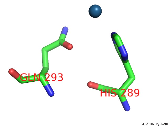
Mono view
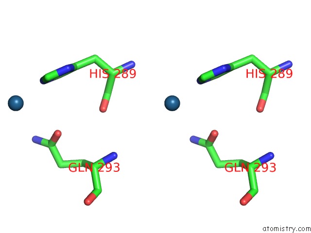
Stereo pair view
Mono view
Stereo pair view
A full contact list of Osmium with other atoms in the Os binding
site number 1 of Crystal Structure of Redox-Inhibited Phosphoribulokinase From Synechococcus Sp. (Strain Pcc 6301), Osmate Derivative within 5.0Å range:
|
Osmium binding site 2 out of 2 in 6hzl
Go back to
Osmium binding site 2 out
of 2 in the Crystal Structure of Redox-Inhibited Phosphoribulokinase From Synechococcus Sp. (Strain Pcc 6301), Osmate Derivative
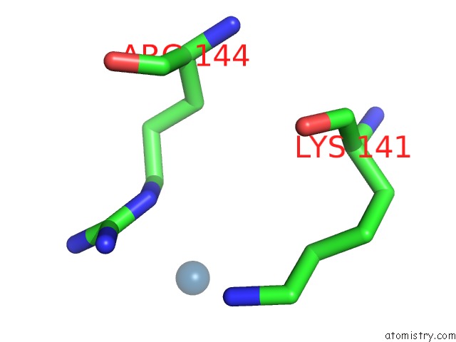
Mono view
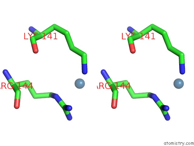
Stereo pair view
Mono view
Stereo pair view
A full contact list of Osmium with other atoms in the Os binding
site number 2 of Crystal Structure of Redox-Inhibited Phosphoribulokinase From Synechococcus Sp. (Strain Pcc 6301), Osmate Derivative within 5.0Å range:
|
Reference:
R.H.Wilson,
M.Hayer-Hartl,
A.Bracher.
Crystal Structure of Phosphoribulokinase From Synechococcus Sp. Strain Pcc 6301. Acta Crystallogr.,Sect.F V. 75 278 2019.
ISSN: ESSN 2053-230X
PubMed: 30950829
DOI: 10.1107/S2053230X19002693
Page generated: Thu Oct 10 09:57:19 2024
ISSN: ESSN 2053-230X
PubMed: 30950829
DOI: 10.1107/S2053230X19002693
Last articles
Zn in 9J0NZn in 9J0O
Zn in 9J0P
Zn in 9FJX
Zn in 9EKB
Zn in 9C0F
Zn in 9CAH
Zn in 9CH0
Zn in 9CH3
Zn in 9CH1